Synapses, depression, and electroconvulsive therapy
- Maarten Laroy
- May 2, 2024
- 2 min read

Updated: May 3, 2024
Electroconvulsive therapy (ECT) is a well-established treatment for late-life depression, but its exact mechanism of action is not fully understood. For my PhD project, I investigated synaptic density changes in patients with depression undergoing ECT using state-of-the-art PET-MR imaging.


The neuroplasticity hypothesis suggests that ECT works by inducing seizures that lead to the formation of new neurons and synapses, crucial for its success. Preclinical research has shown that electroconvulsive stimulation on rodents enhances neurotrophic factors, promotes neurogenesis, and alters synaptic circuitry in the hippocampus. These changes are believed to underlie ECT's mood-improving effects. However, translating these findings from rodents to humans has been challenging.
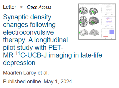
For our study, we recruited patients with depression and conducted brain imaging using a combined PET-MR scanner. Because of this special type of scanner, our patients could receive PET and MRI scanning at the same time, instead of two separate sessions. We then used a specific analysis technique (Voxel-Based Morphometry) to detect within-subject changes in gray matter volume following ECT. In addition, synaptic density was assessed using the novel 11C-UCB-J PET tracer, which targets synaptic vesicle protein 2A (SV2A), a protein that indicates the presence of synapses in brain tissue. The study aimed to explore the relationship between changes in structural gray matter and molecular synaptic density induced by ECT and the treatment's effectiveness.
The analysis revealed significant increases in gray matter in specific brain regions after ECT. However, there were no increases in synaptic density in these regions when looking at the whole group as one. What we did notice was that the changes in synaptic density varied depending on the clinical outcome after ECT, with remitted patients showing an increase in synaptic density and non-remitted patients displaying a small decrease.


This pilot study provides valuable insights into the neuroplastic changes associated with ECT in patients with depression. The findings suggest that while ECT may lead to structural changes in the brain, these changes may not be directly related to synaptic density. The study's small sample size and absence of a control group limit the immediate clinical impact of the findings. Further research with larger sample sizes and longer follow-up periods is needed to validate these findings and explore the link between ECT, neuroplasticity, and therapeutic efficacy.
Our study sheds light on the synaptic density changes following ECT in patients with late-life depression. While the study did not find a direct association between gray matter increase and synaptic density changes, it highlights the potential of PET-MR imaging to investigate neuroplasticity in clinical settings. Further research in this area will contribute to a better understanding of the mechanisms underlying ECT's therapeutic effects and may pave the way for more targeted and effective treatments for depression.
The article can be accessed here
Laroy, M, Vande Casteele, T., Van Cauwenberge, M., Koole, M., Dupont, P., Sunaert, S., Van den Stock, J., Sienaert, P., Van Laere, K., Vandenbulcke, M., Emsell, L., Bouckaert, F. (2024). Synaptic density changes following electroconvulsive therapy: A longitudinal pilot study with PET-MR 11C-UCB-J imaging in late-life depression. BRAIN STIMULATION, in press. doi: 10.1016/j.brs.2024.04.020


